(Microsoft PowerPoint - echo1-1 [\310\243\310\257 \270\360\265\345])
|
|
|
- 하니 만
- 6 years ago
- Views:
Transcription
1 echocardiography RDMS, RT Kang Hye Kyoung
2
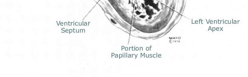
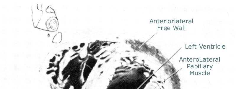
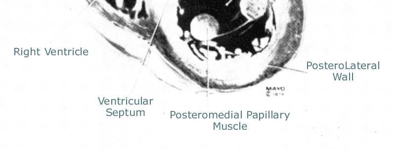
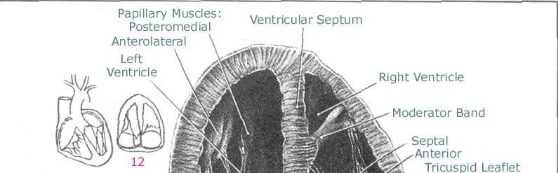
3 Basal surface
4
 심근 (myocardium) 심내막")
5 LAYERS 심막 (pericardieum) visceral layer( 장쪽막 ) + parietal layer( 벽쪽막 ) - pericardial effusion - epicardium( 심외막 ) 심근 (myocardium) 심내막 (endocardium)
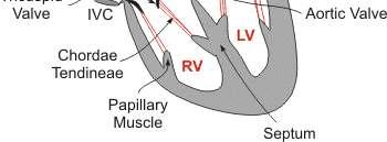
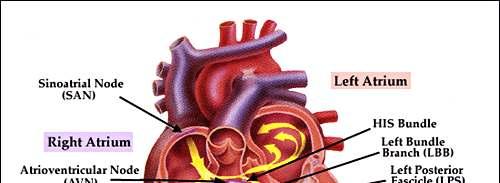
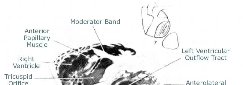
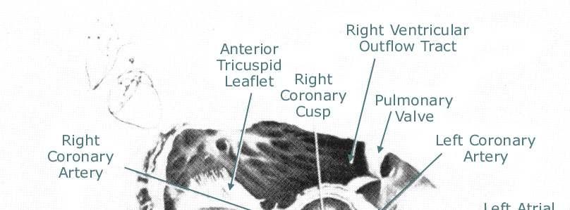
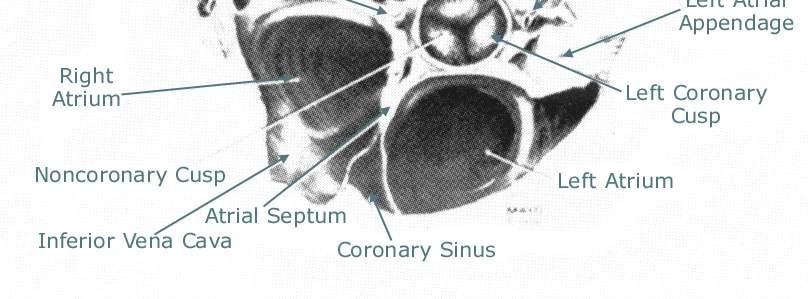
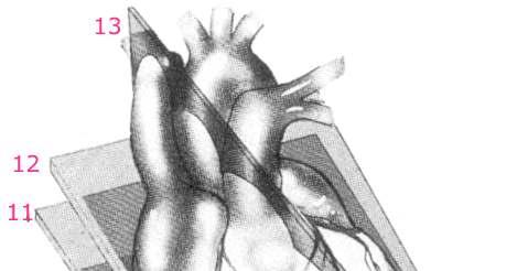
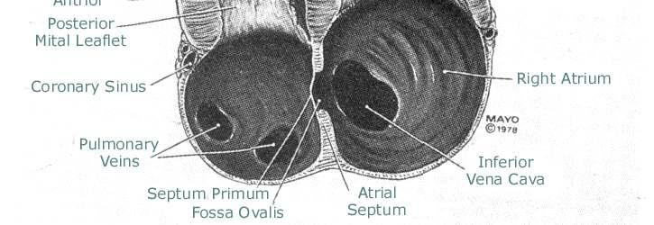
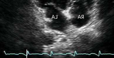
6 Rt atrium
7 Rt ventricle
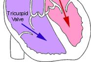
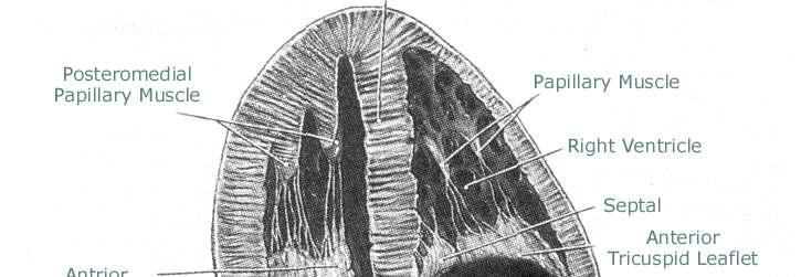
8 Tricuspid valve
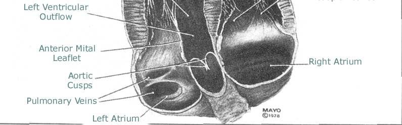
9 Lt atrium
10 Lt ventricle
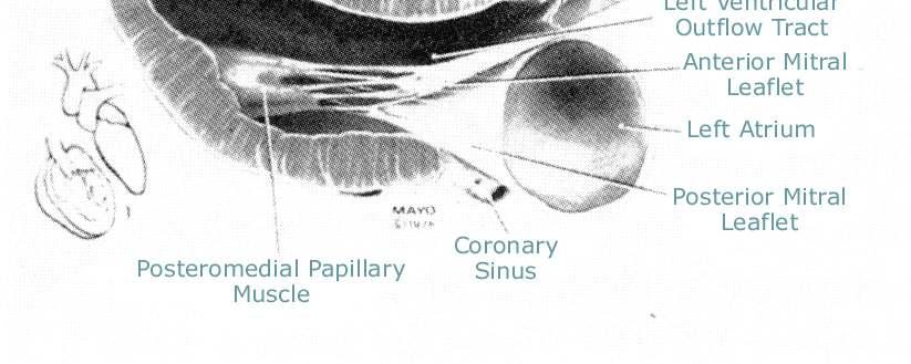
11 Mitral valve
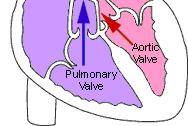
12 Aortic valve
13
14 THE ATRIA AND VENTRICLES 심장은오른쪽 2개, 왼쪽2개 4개의 chamber 우심방, 우심실, 좌심방좌심실 심실을실제내보내는 chamber 수축 ( 심실의수축 ) 할때혈액은심실로부터혈관계를지나다른쪽으로들어올충분한압력으로 eject 심실은수축하고혈액을다시내보내기전에채울 ( 이완할 ) 충분한시간을가져야한다. 심방은채워지는방이다. 심실이수축할때혈액을채운다. 이혈액은이완기에심실을채울수있다.
15 VALVES AND VALVE APPARATUS 판막은심장내에서혈류의방향을조절 심장의효율을극대화하기위해서혈류는한쪽방향으로만흘러야한다. 거꾸로흐르는혈류는펌프로서심장의효율을떨어뜨린다. 사실, 심장은작은양의역류가있어도대부분의혈액은제대로보낸다. 역류하는양이너무많아서감당할수없을때역류하는혈액을받는 chamber은커지기시작한다. 심장에는 4개의판막이있다. 삼첨판, 폐동맥판, 승모판, 대동맥판 방실판막과반월판
16 The Atrioventricular Valves 삼첨판막은오른쪽의방실판막 승모판막은왼쪽의방실판막 AV valve는수축기에심실에서심방으로혈류가역류하는것을막는다. AV valve는 cusps또는 leaflets을갖는다. 혈류정방향으로흐르도록열리고, 거꾸로흐르는것을막기위해닫힌다. 삼첨판막 - Anterior, posterior, septal leaflets 승모판막 - Anterior, posterior AV valve는그기능을돕기위해 apparatus 를갖는다 - chordae tendinae, papillary muscles AV valve apparatus가판막을열거나닫는것은아니다. Valve apparatus의기능은수축기때 AV valve leaflets이탈출 (prolapsing) 하는것을막는다
17 The Semilunar Valves 반월판은심실과이연결되는대혈관사이에위치 폐동맥판막은오른쪽에위치하는반월판 - 우심실과폐동맥사이에위치 대동맥판막은왼쪽에위치 - 좌심실과대동맥사이에위치한다. 반월판은이완기때대혈관에서심실로혈류가역류하는것을막는다. 반월판은정방향의혈류에열리고역방향의혈류에닫히는각각세개의 cusps 또는 leaflets 을갖는다. 폐동맥판막 - Anterior, right posterior, left posterior 대동맥판막 - Right, left, non-coronary : 각각의 cusps 위치에서 Sinus of Valsalva 로알려진 pouch 가있다.
18 Valvular Function Valve 는수동적인구조 심장내에판막을열거나닫는구조물은없다. 판막은심장내압력의변화에의해열리고닫히는것이다.
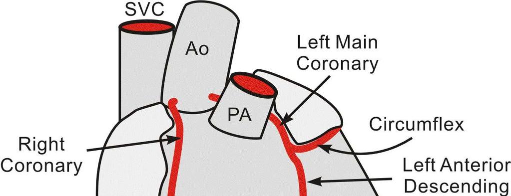
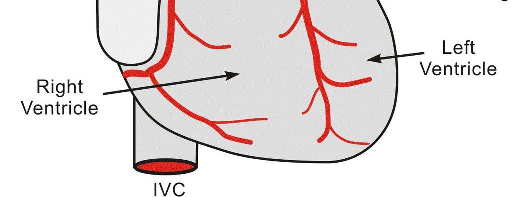
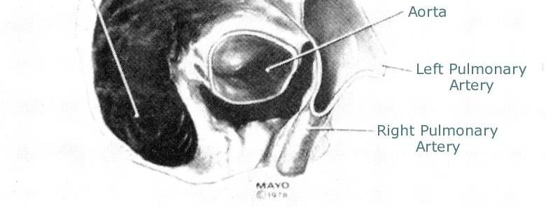
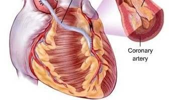
19 CARDIAC BLOOD SUPPLY 심장근육 (myocardium) 은산소의큰 user 혈액은관상동맥 (coronary arteries) 에 myocardium에공급 우, 좌관상동맥 (right and left coronary arteries) 은우, 좌대동맥동 (right and left Sinus of Valslava) 으로부터기시 우관상동맥은우심실은공급하는 acute marginal branch 로갈라진다.- atrioventricular goove를따라심장의후면으로이어지는 posterior descending artery Posterior descending artery는 interventricular groove를따라간다.
20 CARDIAC BLOOD SUPPLY Left coronary artery 는심장의후면에왼쪽 atrioventricular groove 를따라가는 circumflex artery 로이어진다.- left anterior descending coronary artery 가전면 interventricular groove 로간다. 관상동맥은이완기 (diastole) 때혈류를공급받는다. 좌심실이수축할때 (systole) aortic cusp 은 Sinus of Valsalva 를막고열려서관상동맥으로혈류를막는다. 대동맥판막이열릴때 (diastole) 관상동맥에혈류는발생한다.
21
22
23
24
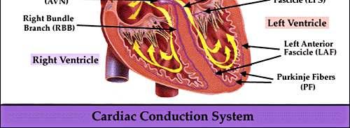
25 CONDUNCTION SYSTEM 심장의기능은폐순환과체순환을통해혈류를내보낸다. 심장의전달체계는심근을전기적으로자극하고, 압력이증가할때수축하게된다. The Sinoatrial Node The Atrioventricular Node The Bundle of His and the Bundle Branches The Purkinje Fibers
26 The Sinoatrial Node( 동방결절 ) SVC 가우심방에들어오는자리에위치 심장의 pacemaker SA node에의해생긴자극이심방에전달되고수축을일으킨다. 자극은 AV node 에전달된다.
27 The Atrioventricular Node 심방에서심실로자극을전달 심방과심실사이에자극을전달하는전기적다리역할
28 The Bundle of His and the Bundle Branches AV node 를떠난자극은 IVS 의오른쪽으로약 1cm 떨어진 common branch 에이르고, 왼쪽과오른쪽으로나뉜다. 이 branch 가심첨부까지자극을운반
29 The Purkinje Fibers 심근에자극을보내는섬유조직 자극은심내막 (endocardium) 에서심근 (myocardium) 을거쳐심외막 (epicardium) 으로전달
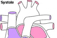
30 Systolic 대동맥과폐동맥으로혈액을방출하는심실수축 대개 0.3~0.4 초동안지속되는심실수축에뒤이어방출기 ( 放出期 ) 가시작되는데, 그동안 80~100 cm3의혈액이심실에서방출 수축기동안동맥혈압은최고에도달 ( 수축기혈압 ) 정상적으로는 120 mm Hg 혈관벽의팽창성때문에심실의압력보다약간낮다. 심방수축기는심실이다시혈액으로채워지는심실확장기끝무렵에시작된다.
31 Diastolic 두수축기사이에일어난다. 이완기는 0.5~0.6초동안지속되는데그동안급속히혈액이채워지고 ( 차단되었던혈액이동맥으로부터밀려들어오면서 ), 뒤이어잠깐동안심방수축이일어난다. 동맥의혈압 - 80mmHg
32
33 Diastolic valve
34 Systolic valve
35
36
37
38 Electrophysiology 탈분극 (Depolarization) 전기적자극을받아파동이일어남, 심장은수축한다. 재분극 (repolarization) 제자리로돌아옴
 T-wave")
39 Electrophysiology P-wave 심방수축 (SA node) QRS complex 심실수축 (AV node~bundle branchs) T-wave 심실재분극
40 심장초음파검사법 경흉부심초음파검사 경식도심초음파검사 조영심초음파 bubble이혈류에들어가면부옇게떠다니는특징을이용한검사법으로좌우단락을확진하거나심근허혈의연구에이용 부하심초음파 약물및운동부하를주면서심초음파검사를하여관상동맥질환의진단과심기능의평가에이용 혈관또는심장내초음파검사 심도자 (cather) 에탐촉자를부착하여여러혈관및심장내강에넣어검사하는방법으로동맥경화증및중재적시술의합병증판단에사용
41 TTE(Trans Thoracic Echo.) Echo exam
42 TTE(Trans Thoracic Echo.) 1. Two-dimensional echocardiography (2D) 2. M-mode echocardiography 3. Doppler echocardiography - Color Doppler - Pulsed wave (PW) Doppler - Continuous wave (CW) Doppler
43 M-mode echocardiography 하나의선만으로초음파를전송및수신을하기때문에움직이는구조물을기록하는데 2D 보다민감도가높다. 시간별로반사되는깊이와강도가그래프로그려진다. 판막의열림, 닫힘, 심실의움직임과같은운동의변화를표시할수있다.
44 M-mode echocardiography
45 Pulsed wave (PW) Doppler 국소적인흐름장애나어느작은부분에서혈액의속도를측정 Sample volum의위치에서특정부분의혈류속도측정 시간지연으로샘플링할수있는비율이한정되기때문에측정할수있는최대속도는약 2m/s 이다. Aliasing 현상주의
46 Continuous wave (CW) Doppler 지속적인송신과수신이가능하다. 빠른속도를측정하는데유용 각신호가어느지점에서발행하는지알수없다.
47 용도 2D : 해부, 심실과판막의움직임, M-mode 와도플러초음파를위한위치선정 M-mode : 치수의측정, 특정부위의시간측정 Pulsed wave Doppler : 정상적인판막흐름의패턴, 좌심실이완기능, 일회박출량과심장박출량 CW Doppler : 판막협착증의중증도, 판막역류의중증도
48 초음파를이용한심질환의진단 심실기능의평가 호흡곤란과심부전의평가 심잡음 비특이적인심전도 흉통 심장에서기인한색전 저혈압, 심장성쇼크 심장좌상및공여자심장평가 고혈압
49 제한점 과도비만환자 가슴이기형인환자 만성폐질환자 ( 만성환기장애, 폐과팽창, 폐섬유화증 )
50 심실기능의평가 좌심실의기능을이해하는것은모든심장검사에서기본적인단계 심초음파를이용한정량적인평가는환자의증상을설명, 적절한치료선택, 적절한수술시기결정 환자의수축기와이완기능평가
51 호흡곤란과심부전의평가 호흡곤란은비정상적인질환의임상양상 수축, 이완기능평가 판막의구조와기능평가 심장단락평가 중심정맥과판막의호흡변형평가 심근증여부 폐동맥고혈압 기타
52 비특이적인심전도 심전도는몇몇심장의병적상태를진단에매우유용 그러나많은심전도이상소견은특이적이거나진단적이지못하다. 심전도이상은수술전평가에있어서관심이증대 비특이적심전도이상은수술적처치나중재적시술후에도발생할수있다. 이와관련된심근허혈이나심근경색등에대한평가에심초음파가이용
53 비특이적인심전도 심근경색과허혈에의한벽운동평가 Pericardial effusion Intracardiac mass Pulmonary embolism LV hypertrophy Valvular heart disease
54 흉통 심전도이상이없으며증상이있을때유용 Ischemia or infaction Aortic dissection Pericarditis Valvular heart disease Cardiomyopathy Pulmonary embolism mass
55 심장에서기인한색전 심장원인 - 심내혈전 - 종양 - Vegetation - 심방중격류 - Foramen ovale를통한 embolism - 대동맥의 ulcerated plaques
56 저혈압, 심장성쇼크 환자곁에서시행하는가장의의있는진단도구 LV and RV function Valvular abnormalities Cardiac tamponade LV outflow tract obstruction Hypovolemia(small LV cavity size with hypercontractile walls) Pulmonary embolism Cardiac shunt Aortic dissection Cardiac rupture
57 고혈압 LV cavity and wall dimensions LV mass index LV systolic and diastolic function Dynamic LV outflow tract obstruction
58 Cardiac condition and their associated symptoms Congestive heart failure LV failure tachycardia fatigue with exertion dyspnea with mild exercise (due to pulmonary edema) paroxymal nocturnal dyspnea with cough cough (in advance CHF) with occasional hemoptysis cyanosis (central)
59 RV failure increasing fatigue edema beginning at ankles ascites in later stages weight gain secondary to retained fluid cyanosis (peripheral)
60 Acute Pulmonary Edema (secondary to LV failure) Extreme dyspnea Cyanosis Restlessness and anxiety hemoptysis
61 Superior Vena Cava Syndrome Obstruction of the SVC, often a lung tumor Edema of the upper extremities, face and neck
62 Mitral Valve Prolapse Usually asymptomatic Chest pain Palpitations Fatigue dyspnea
63 Mitral Insufficiency Fatigue Dyspnea Congestive heart failure
64 Mitral stenosis Exertional dyspnea Late paroximal nocturnal dyspnea ( 발작성야간호흡곤란 ) Exertional tachycardia Pulmonary edema (late) Right heart failure (late)
65 Aortic Insufficiency Asymptomatic if mild Dyspnea Fatigue on exertion Syncope Chest pain Congestive heart failure (late)
66 Aortic Stenosis Fatigue Exertional dyspnea Angina (inadequate coronary perfusion) Syncope on exertion
67 Tricuspid Insufficiency Neck vein distension Peripheral edema Abdominal distension (ascites)
68 Tricuspid stenosis Neck vein distension Peripheral edema Abdominal distension (ascites)
69 Pulmonary Insufficiency Few specific symptoms May be secondary to pulmonary hypertension
70 Pulmonary Stenosis Pulmonic stenosis almost always a congenital lesion possibly with associated abnormalities symptoms will vary
71 IHSS (idiopathic hypertrophic subaortic stenosis) Patient may be asymptomatic Fatigue Dyspnea Palpitations Precordial( 전흉부의 ) discomfort or angina-type chest pain Syncope Sudden death
72 Myxoma Fever Weigth loss Congestive heart failure Syncope Symtoms may clinically mimic mitral stenosis due to tumor prolapse through the mitral valve
73 Pulmonary Embolism( 폐색전증 ) 폐동맥이나그가지가혈류에의해운반된혈병또는이물에의해폐쇄 경색증을일으키는질환 대개는정맥혈전, 특히다리의심재정맥혈전이폐동맥에유입됨으로인해폐동맥이폐색되어폐혈관계의순환이저해되는질환 외상특히고령자의골반이나대퇴골의골절 화상 수술 산욕 심장질환 악성종양 혈액질환등의경우에발생하기쉽다
74 Dyspnea of sudden onset Retrosternal pain Anxiety and restlessness Pallor( 창백 ) with peripheral cyanosis Hemoptysis( 객혈 )
75 Pulmonary Hypertension Decreased exercise tolerance with syncope on exertion Fatigue and weakness Progressive dyspnea RV failure
76 Acute MI May be silent (asymptomatic) Chest pain (similar to angina but more severe)with squeezing or pressure radiating to jaw( 턱 ) and left arm Restlessness and apprehension( 불안감 ) dyspnea
77 Aortic Dissection Back or chest pain (excruciating) of sudden onset Some patients may be asymptomatic
78 Hypertension May be asymptomatic Dizziness Fatigue May have symtoms of LV failure in May have symtoms of LV failure in chronic hypertension
79 syncope Hypotension
80 Pericarditis Sharp left-sided pain aggravated by deep breathing, swallowing, coughing and movement. Pain is relieved by sitting up and leaning forward. Fever and chills( 오한 ) Weakness Cough
81 Endocarditis Fever and chills Malaise ( 불안, 권태감 ) Arthralgia ( 관절통, painful joints)


82 Standard Image Plane of 2-D
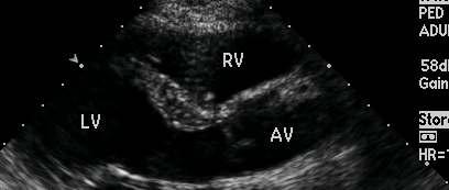
83 Standard Image Plane of 2-D Parasternal long axis view Parasternal short axis view Apical view 4 chamber view 2 chamber view 3 or 5 chamber view Subxiphoid view / Suprasternal view
84

85 Anatomy of the PLAX View


86 2D Echo. : PLAX view Parasternal Long Axis View
87
88 LV and LA Measurements LV posterior wall (D) Normal Values Male Left atrium 3.0 ~ 4.5 LV diastolic dimension 4.3 ~ 5.9 LV systolic dimension 2.6 ~ 4.0 LV Interventricular septum (D) 0.6 ~ 1.3 Aortic Measurements Aortic annulus Sinus of Valsalva Sinotubular junction Aortic arch Abdominal aorta 0.6 ~ 1.2 Absolute value 1.7 ~ ~ ~ ~ ~ 2.2 Female 2.7 ~ ~ ~ ~ ~ 1.1 Indexed to BSA 1.1 ~ ~ ~ ~ ~ 1.3
89
90
91
92

93 2D Echo. : PSAX view
94
95
96

97 2D Echo. : PSAX view
98
99
100

101 2D Echo. : PSAX view
102
103

104 2D Echo. : Apical view

105
 3. Mitral Annulus Velocity ( E ) 4.")
106 Apical 4 chamber view 1. MV, TV color Doppler 2. Mitral inflow 측정 ( E, A wave, DT ) 3. Mitral Annulus Velocity ( E ) 4. Wall motion analysis 5. MR, TR velocity 측정 6. RV
107

108 2D Echo. : Apical view
109
110 Apical 3 chamber view 1. AV & LVOT 관찰 2. Wall motion analysis - Anteroseptum - Post. wall
111

112 Apical 2 chamber view 1. MV 정밀관찰 ( MVP ) 2. Wall motion analysis - Inf. wall - Ant. wall

113 2D Echo. : Suprastenal view


114 2D Echo. : Subcostal view


115 Subcostal view 1. IVC plethora 관찰 2. Atrial septum - ASD
116 심장초음파검사 심초음파도를이용한심장내경의측정 좌심실의수축기능평가 좌심실의이완기능평가및이완기심부전 혈역학적평가 심초음파를이용한관동맥혈류관찰과예비력평가 판막질환
117 심장초음파검사 감염성심내막염 허혈성심질환 심근병증 심낭질환
118 심초음파도를이용한심장내경의측정 M mode 심초음파도측정 2D 심초음파도측정 심실기능평가
119 2D

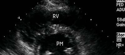
120 M mode : LV mid level Parasternal Short Axis View papillary Muscle Level 에서그림과같이 M-mode 실행

121 M mode LV EF(M-mode) 측정방법
122 혈역학적평가 1 회박출량과심박출량 압력차 판막면적 심장내압력 도플러심장초음파
123 Stroke volume(sv) 1 회심박출량 수축기에좌심실에서보내지는혈류량 Stroke volume = (end-diastolic volume) (end-systolic volume) 정상범위 75~100ml 구멍을가로지르는흐름은단면적과유속을곱한값이다 흐름 = 단면적 x 속도
124 Ejection fraction(ef) 박출량 수축기에박출한이완기말좌심실용적의백분율 Ejection fraction = stroke volume/(enddiastolic volume) 정상범위 > 55%
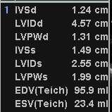
125 Ventricular systolic function 2D, M-mode 측정 EF(%) =EDV-ESV/EDV*100% 55-80% FS(%)=LVIDd-LVIDs/LVIDd*100% 25-45% SV=EDV-ESV LVES-LVED-LV end-diastolic dimension LV end-systolic dimension EDV=LV end diastolic volume ESV=LV end systolic volume
126 LV 용적과 EF 측정 1. Cube method V=D³ 2. Teichholz method V=[7/(2.4+D)]XD³ 3. Simpson s method, Area-length method
127 Modified Simpson Method
128 Cardiac output 단위시간당 (1 분 ) 좌심실에서내보낸혈류량 Cardiac output = stroke volume x heart rate 정상범위 5.0~7.5 liters/min


129 Abnormal pattern M-Mode Mode Ld Ls
130 Parasternal Long Axis 1. MV, AV 의 Function ( Color Doppler ) 2. LV, Aorta, LA Size 측정 (Chamber size) 3. LVH, pericardium 관찰 4. Septum, Post wall 의 wall motion 측정
131 Parasternal Short Axis 1. Aortic valve level - PV PW and AV, TV color doppler - LA, AV size 측정 ( M- mode ) 2. Mitral Valve level - MV morphology 관찰 ( M-mode) 3. Mid LV level ( Papillary muscle level ) - LV EF 측정 ( M-mode ) - Wall motion 관찰 4. Apex level - Wall motion 관찰 - Wall motion 관찰

132 M mode : AV level Adult Normal value Ao root diameter : 21~37mm LA diameter : 22~40mm Parasternal Short Axis View Aortic valve Level에서그림과같이 M-mode 실행

133 Interpretation of Report LV dimension LV wall thickness LV systolic function RV dimension IVC plethora LA and Aortic size Pericardial effusion E-point septal separation
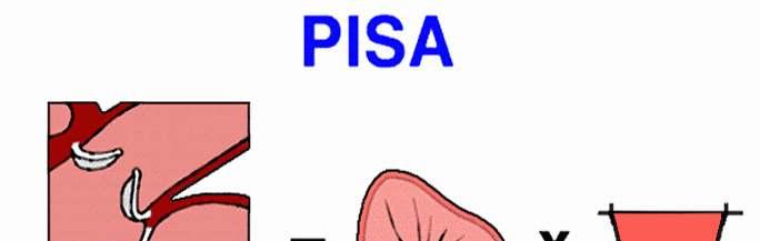
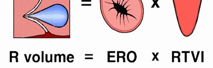
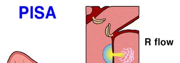
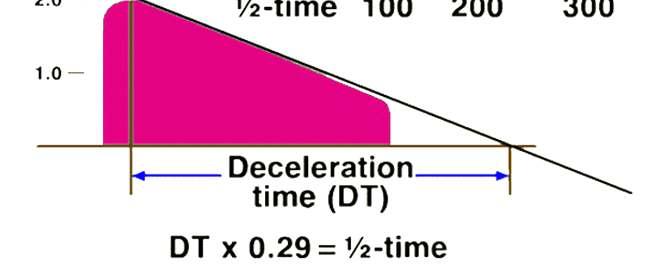
134 좌심실의이완기능평가및이완기심부전 정상이완기능 : 심방의압력증가없이심실이적절히채워짐 평가방법 - E/A ratio : 유입혈류최고속도 (E velocity), 심방수축에의한후기혈류속도 (A velocity) 의비 E/A = 1~2 - DT time : 감속시간으로이완기능장애시증가 160~240ms
135 Assessment of Diastolic Function : MV PW & MV anntdi Normal Pattern PW Doppler Tissue Doppler
136 Assessment of Diastolic Function Sohn DW et al, J Am Coll Cardiol 1997;30:474) Sohn DW et al, J Am Coll Cardiol 1997;30:474)
137 Pulsed Wave Doppler : MV, Pvein and PV Normal Pattern MV - PW Doppler Normal : PVs2>PVd PV PW Doppler Impaired : PVs2>>PVd Pseudopattern : PVs2<PVd Restrictive : PVs2<<PVd
138 Interpretation of Report Valve morphology Represent LV diastolic function Represent severity of mitral stenosis Presence and severity of mitral regurgitation Echo score in mitral stenosis
139 Normal Values Normal ranges for measures of systolic and diastolic function 2D Measurement Fractional shortening (%) End-diastolic volume (ml) End-systolic volume (ml) Ejection fraction (%) Doppler 28 ~ ~ 166 (male) 3 ~ 67 (male) 50 ~ ~ 129 (female) 9 ~ 57 (female) Systolic velocity integral 15 ~ ~ 25 (elderly) Mitral E velocity (cm/sec) 44 ~ ~ 90 (elderly) Mitral A velocity (cm/sec) 20 ~ ~ 87 (elderly) E/A ratio 0.7 ~ ~ 1.7 (elderly) Time interval Mitral deceleration time (msec) 139 ~ ~ 282 (elderly) Isovolumic relaxation time(msec) 54 ~ ~ 124 (elderly)
140 Valvular Heart Disease MR(mitral regurgitation) MS(mitral stenosis) AR(aortic regurgitation) AS(aortic stenosis)
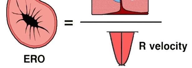
141 Diagnostic Evaluation of MR 1. Mitral valve anatomy 1) Cause of regurgitation 2) Normal, excessive, or restrictive leaflet motion 3) Reparability of valve 2. LV size and systolic function 1) Dimensions and volumes 2) Ejection fraction(ef) 3) Regional wall motion 3. LA size 4. Pulmonary artery pressure & RV function 5. Regurgitant severity 1) Color Flow Doppler 2) CW Doppler signal strength and shape 3) Regurgitant volume(rv) 4) Regurgitant orifice area (ERO)
 view 모두관찰하는것이바람직하다.")
 mild Apical 4chamber View")
142 MR(Mitral Valve regurgitation) PLAX view or Apical 4ch(2ch) view 모두관찰하는것이바람직하다. 보통 MR flow 가 LA 에미치는정도에 따라서약한정도부터 trivial(minimal) mild Apical 4chamber View moderate -severe 로구별한다.

143 Color Doppler 로측정한 MR 의중증도 Color Flow Mapping +1 = trivial +2 = mild +3 = moderate +4 = severe
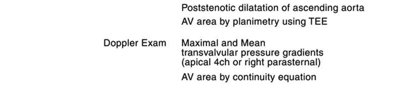
144 PISA method 로측정한 MR 의중증도 Proximal Isovelocity Surface Area method Proximal Convergence Method Proximal Side regurgitant orifice (r) r 42cm/2 (V o ) (v)
145
146


147 PISA method Process Acquisition Review & Analysis ZOOM Change Baseline


148 PISA method Process Acquisition Review & Analysis Measure Pisa
149 PISA method 로측정한 MR 의중증도 Estimation of Mitral Regurgitation severity by ERO: Grade I: < 0.10 mm 2 Grade II: mm 2 Grade III: mm 2 Grade IV: >35mm 2 Estimation of Mitral Regurgitation severity by RV(Regurgitant Volume) Grade I = 1-25cc Grade II = 25-40cc Grade III =40-55cc Grade IV = >55cc
150 MS(Mitral Valve Stenosis)


151 MS(Mitral Valve Stenosis) 2D and Color Doppler


152 MS(Mitral Valve Stenosis) CW Doppler

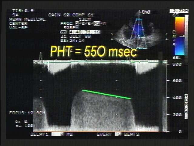
153 MVA(Mitral Valve Area) 2D and CW Doppler 1. 2D Planimetry : MV area trace 2. Doppler : MVA=220/PHT MVA Normal : 4-6cm2 Mild : cm2 Severe : < 1.0 cm2
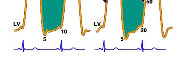
154 Mitral Stenosis Severity MVA Mean PG CO (cm 2 ) (mmhg) (L/min) Mild 1.5~ ~9.0 Moderate 1.0~1.5 5~10 5.5~6.5 Severe ~5.0

155 Mitral Valve Area
156 EchoCG in MS Mitral Stenosis Measurement of MVA Planimetry and PHT Morphologic evaluation of MV; Echo score PMV vs. MVR LA thrombi? TEE > TTE Combined MR? Pulmonary hypertension?
157 Mitral Stenosis Echo score Mobility Thickening Calcium Subvalve Mobile valve Thin No bright echo Spars echo Immobile valve Severely thickened Multiple bright echo Multiple thick chordae
158 PMV ; Contraindications High echo score Mitral Stenosis Good Intermediate Poor LA thrombi Mural thrombi; absolute CIx Appendageal thrombi; relative CIx Moderate to severe MR Combined aortic valve disease
159 AS(Aortic Valve Stenosis)
160 AS 의특징 Increased thickeness of AoV leaflet LVH and increased LV mass Decreased LV systolic function(late) LAE

161 Aortic Stenosis
162 Aortic Stenosis

163 Aortic Stenosis
164 Aortic Stenosis Continuity equation for AVA
165 Aortic Stenosis Severity Mild Moderate Severe Very severe AVA (cm 2 ) > ~ ~1.0 <0.75 AVA index (cm 2 / M 2 ) > ~ ~0.6 <0.4
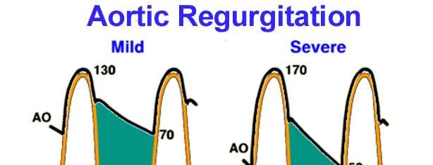
166 Aortic Regurgitation mmhg Mild AR BP 140/70 Severe AR BP 140/40 50 LVEDP 14mmHg LVEDP 28mmHg

167 Aortic Regurgitation 150 Mild AR Severe AR
168 Aortic Regurgitation Aortic valve regurgitant(ar)


169 Aortic Valve Area AVA Normal : cm2 Mild : cm2 Moderate : Severe : < 0.75 cm2
170 Aortic valve regurgitant(ar) Aortic valve regurgitant(ar)- Ao Arch view
171 Interpretation of Report Valve morphology Presence and severity of aortic regurgitation Presence and severity of aortic stenosis
172 TR(Tricuspid Valve Regurgitation)


173 TR method 로측정한 pulm. HT PASP=RVSP PASP =RAP+TRp
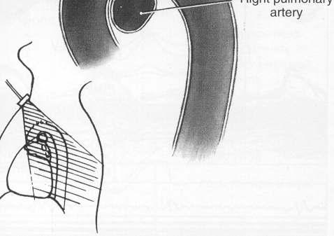
174 TR method 로측정한 pulm. HT Normal PASP=18-25 mmhg Mild pulm. HT =30-40 mmhg Moderate pulm. HT=40-70 mmhg Seevere pulm. HT=>70mmHg
175 Interpretation of Report Valve morphology Presence and severity of tricuspid regurgitation Presence and severity of pulmonary hypertension Tricuspid inflow


176 Interpretation of Report M-mode and 2D Measurements Valve functions
177 감염성심내막염
178 감염성심내막염
179 감염성심내막염
180 ASD
181 ASD
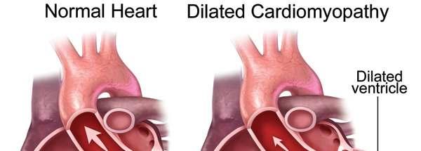
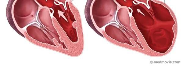
182 ASD
183 AS
184 Bicuspid valve
185 aorta
186 Aortic arch
187 Pulmonary artery
188 myxoma
189 심초음파를이용한관동맥혈류 관찰과예비력평가
190
191
192 Ischemic heart disease
193
194 Dilated cardiomyopathy
195 Dilated cardiomyopathy
196
197 Hypertrophic cardiomyopathy
198 Hypertrophic cardiomyopathy
199 심낭질환
200 허혈성심질환
201 Prosthetic valve
202 Prosthetic valve
203 Prosthetic valve
204 Prosthetic valve
205 ASD
206 ASD
207 ASD
208
<4D F736F F F696E74202D20BFA1C4DA5FC0D3BBF3C3CAC0BDC6C42E BC8A3C8AF20B8F0B5E55D>
 심장 (Heart) 심초음파 서울대병원내과 김용진 심장은평생몇번이나뛸까요? 1) 3 백만번 2) 3 천만번 3) 3 억번 4) 30 억번 30 억박동, 2 억리터의혈액 Cause 심장질환 6.2 12.4 11.1 16.2 뇌졸중 4.3 8.5 7.7 11.3 기타심혈관질환 2.6 5.1 6.0 8.8 심혈관질환 13.1 26.0 24.8 36.3 총사망 50.4
슬라이드 제목 없음
 심장영상진단학 세종병원영상의학과김양민 X - 선의발견 At 1895, Wilhelm Conrad Roentgen, a German scientist, discovered X-rays... - 2010 Perfusionist symposium - X- 선의특성 빛과같은전자기파의일종 렌즈에통하여도굴절되지않는다 거울이나금속면에서반사되지않는다 물질을직선으로투과한다
<B0E6C8F1B4EBB3BBB0FA20C0D3BBF3B0ADC1C E687770>
 심전도연수강좌 : 처음시작하는사람들을위한심전도 연세대학교원주의과대학순환기내과학교실 안민수 Cardiac Electrophysiology I : Automaticity : 60-100 회 /min, His bundle : 40-60 회 /min Bundle branch : 20-40 회 /min Purkinje fiber : 20 회 /min Cardiac Electrophysiology
심장의구조와기능 (2 of 2) 심장판막 : 혈액을한방향으로만흐르게하는판막방실판막 (Atrioventricular or AV valves 삼첨판 (Tricuspid valve) 이첨판 (Bicuspid valve or mitral valve) 동맥판막 : - 폐동맥판
 심장판막 : 혈액을한방향으로만흐르게하는판막방실판막 (Atrioventricular or AV valves 삼첨판 (Tricuspid valve) 이첨판 (Bicuspid valve or mitral valve) 동맥판막 : - 폐동맥판") 심장의구조와기능 (1 of 2) 기능 : 근육성펌프 ; 폐를통과하여말초조직으로순환시킨다. 심장병 (Heart disease): 심장펌프의기능장애 - 우심장 : 폐순환을담당 - 좌심장 : 체순환을담당 심장의구조와기능 (2 of 2) 심장판막 : 혈액을한방향으로만흐르게하는판막방실판막 (Atrioventricular or AV valves 삼첨판 (Tricuspid
심장의구조와기능 (1 of 2) 기능 : 근육성펌프 ; 폐를통과하여말초조직으로순환시킨다. 심장병 (Heart disease): 심장펌프의기능장애 - 우심장 : 폐순환을담당 - 좌심장 : 체순환을담당 심장의구조와기능 (2 of 2) 심장판막 : 혈액을한방향으로만흐르게하는판막방실판막 (Atrioventricular or AV valves 삼첨판 (Tricuspid
Anatomy and Physiology of Heart OCW4 1
 Anatomy and Physiology of Heart OCW4 1 학습목표 학습자는해부생리학과목에서학습한심장의기본구조와기능을복습함으로서, 1) 자발적으로요약설명할수있다. 2) 심박출량에영향을주는요인을규명할수있다. 2 심장의기본구조와생리 심장무게 : 약 300그램 심장크기 : 자신의주먹정도 주요구성조직 : 근육조직 ( 심근, 유두근 ) 위치 ; 종격동의중앙,
슬라이드 1
 임상생리학 ( 심장초음파 )-4 2-D 심장초음파검사및대표적이상소견 Konyang University Dept. Biomedical Laboratory Science Keun-Sik Kim Ph.D 주요내용및활동 2-D 의개념및심장의해부학적단면? ( 사전지식공유자유토론 ) 수업방법 토론식 국시관련문제제시및토론 ( 자료공유 ) 토론식 2-D 심장초음파강의 국시관련문제제시및해결
 Fluoroscopic anatomy for supraventricular tachycardia ablation Hui-Nam Pak, MD, PhD Division of Cardiology, Yonsei Cariovascular Center and Cardiovascular Research Institute, Yonsei University College
부정맥 (Cardiac Arrhythmias, Dysrhythmias) ECG 는 atrium 에서 ventricle 로즉 (SA node AV node His bundle bundle branch Purkinje fiber) 의 normal route 를따라 depo
 ECG 는 atrium 에서 ventricle 로즉 (SA node AV node His bundle bundle branch Purkinje fiber) 의 normal route 를따라 depo") 부정맥 (Cardiac Arrhythmias, Dysrhythmias) ECG 는 atrium 에서 ventricle 로즉 (SA node AV node His bundle bundle branch Purkinje fiber) 의 normal route 를따라 depolarization 하기만하면 P wave 와 QRS complex 는 normal appearance
부정맥 (Cardiac Arrhythmias, Dysrhythmias) ECG 는 atrium 에서 ventricle 로즉 (SA node AV node His bundle bundle branch Purkinje fiber) 의 normal route 를따라 depolarization 하기만하면 P wave 와 QRS complex 는 normal appearance
Jksvs019(8-15).hwp
.hwp") Grade I Grade II Grade III 12 대한혈관외과학회지 : 제 20 권 제 1 호 2004 Control Group A Group B Fig. 4. Microscopic findings of vein wall in control, group A and group B on the day of 7 after venous occlusion. The
Grade I Grade II Grade III 12 대한혈관외과학회지 : 제 20 권 제 1 호 2004 Control Group A Group B Fig. 4. Microscopic findings of vein wall in control, group A and group B on the day of 7 after venous occlusion. The
ºÎÁ¤¸ÆV10N³»Áö
 A case of nonischemic dilated cardiomyopathy with ventricular tachycardia treated by ICD ABSTRACT Dilated cardiomyopathy (DCM) is a syndrome characterized by left or biventricular dilatation and impaired
<4D F736F F F696E74202D20B0B3BFF8C0C7BFACBCF6B0ADC1C220B0ADC0C7B7CF5FC1B6B1B8BFB5>
 순환기내과 조구영 Disorder causing dyspnea Pulmonary Air flow limitation, restrictive, chest wall, pulmonary circulation Cardiac Coronary Valvular Myocardial: systolic or diastolic disorder Anemia Peripheral circulation
Microsoft PowerPoint - 2- 남기병
 쉽게풀어보는심전도 울산대서울아산병원남기병 심전도진단 Chamber enlargement (hypertrophy) Bundle branch block Myocardial infarction WPW ST-T change: Nonsp ST-T change, Early repolarization Arrhythmia: tachycardia, PVC, AF 남자 63
Case Reports Korean Circulation J 심한석회화를동반한거대 Valsalva 동동맥류 류제영 정명호 강경태 이상현 박종철 안영근김윤현 조정관 안병희 김상형 박종춘 강정채 A Giant Aneurysm of the S
 Case Reports Korean Circulation J 2001311 114-118 심한석회화를동반한거대 Valsalva 동동맥류 류제영 정명호 강경태 이상현 박종철 안영근김윤현 조정관 안병희 김상형 박종춘 강정채 A Giant Aneurysm of the Sinus of Valsalva with Calcification Jay Young Rhew, MD,
ºÎÁ¤¸ÆV10N³»Áö
 A case of atrioventricular dissociation with interference ABSTRACT Interference dissociation is one of the most interesting arrhythmias. Once thought to be a rare arrhythmia, it is now considered a very
06. Interpretation of diagnostic test 521.hwp
 대한내과학회지 : 제 92 권제 6 호 2017 https://doi.org/10.3904/kjm.2017.92.6.521 Interpretation of diagnostic test 심초음파를이용한좌심실이완기능의평가 1 가천대길병원심장내과, 2 가천대학교의과대학심장내과 최하늘 1 ㆍ신미승 1,2 Echocardiographic Evaluation of Left
 ECG & EP CASES Young-Keun On, MD, PhD Division of Cardiology, Department of Medicine Cardiac & Vascular Center, Samsung Medical Center Sungkyunkwan University School of Medicine, Seoul, Korea A case of
untitled
 Korean J Clin Lab Sci. 2019;51(2):270-275 https://doi.org/10.15324/kjcls.2019.51.2.270 Korean Society for Clinical Laboratory Science CASE REPORT Assessment of the Severity of Degenerative Aortic Stenosis:
Case Reports Korean Circulation J 1999;29 10 : 흉부둔외상후 8 년뒤에증상이발현된삼첨판폐쇄부전 1 례 김연중 문건식 김재성 황흥곤 Tricuspid Insufficiency Detected 8 Years Later F
 Case Reports Korean Circulation J 1999;2910:1133-1137 흉부둔외상후 8 년뒤에증상이발현된삼첨판폐쇄부전 1 례 김연중 문건식 김재성 황흥곤 Tricuspid Insufficiency Detected 8 Years Later Following a lunt Chest Trauma Yeoun Jung Kim, MD, Keon
ºÎÁ¤¸ÆV10N³»Áö
 case of brugada syndrome presented as chest pain STRCT rugada syndrome was described in 1992 as a new clinical entity characterized by electrocardiographic STsegment elevation in the right precordial leads
(Microsoft PowerPoint - S13-3_\261\350\273\363\307\366 [\310\243\310\257 \270\360\265\345])
![(Microsoft PowerPoint - S13-3_\261\350\273\363\307\366 [\310\243\310\257 \270\360\265\345])](/thumbs/100/144769741.jpg "(Microsoft PowerPoint - S13-3_\261\350\273\363\307\366 [\310\243\310\257 \270\360\265\345])") Cardiovascular Disease in Metabolic Syndrome 김상현 보라매병원내과 서울대학교의과대학내과학교실 Contents Metabolic syndrome and Cardiovascular system CVD Mortality Coronary artery disease Heart failure Atrial fibrillation Management
Cardiovascular Disease in Metabolic Syndrome 김상현 보라매병원내과 서울대학교의과대학내과학교실 Contents Metabolic syndrome and Cardiovascular system CVD Mortality Coronary artery disease Heart failure Atrial fibrillation Management
untitled
 대흉외지 2010;43:58-62 DOI:10.5090/kjtcs.2010.43.1.58 증례보고 반회전동맥간전환술후양심실유출로의중단기변화 2 예보고 김정원 * ㆍ조준용 * ㆍ김근직 * ㆍ이종태 * ㆍ김규태 * Changes of the Biventricular Outflow Tract after a Half Turned Truncal Switch Operation
Original Articles 승모판협착증환자에서심초음파로측정한 Abstract 승모판막넓이의정확성 : 수술중측정한 승모판막넓이와의비교 * 한창엽 김기식 한성욱 허승호 배장호 김윤년 김권배 Accuracy of Mitral Valve Area in
 Original Articles 28 2 1998 승모판협착증환자에서심초음파로측정한 Abstract 승모판막넓이의정확성 : 수술중측정한 승모판막넓이와의비교 * 한창엽 김기식 한성욱 허승호 배장호 김윤년 김권배 Accuracy of Mitral Valve Area in Patients with Mitral Stenosis Measured by EchocardiographyCompared
<313520C1F5B7CA B0ADB9CEB1D42DB9DAC1A4B6FB D E687770>
 대한내과학회지 : 제 82 권제 5 호 2012 http://dx.doi.org/10.3904/kjm.2012.82.5.609 영구형인공심박동기삽입술후발생한스트레스성심장근육병증 1예 경상대학교의학전문대학원내과학교실 강민규 김나영 박정랑 황석재 박용휘 곽충환 황진용 Stress-Induced Cardiomyopathy as a Complication of Permanent
Case Reports Korean Circulation J 2000;30 10 : Urokinase 정맥주사로치료한재발성폐혈전색전증 1 예 박경창 김지수 김삼 육청미 이상무 정성원 이남호 박대균 Recurrent Pulmonary Thromboembo
 Case Reports Korean Circulation J 2000;3010:1285-1290 Urokinase 정맥주사로치료한재발성폐혈전색전증 1 예 박경창 김지수 김삼 육청미 이상무 정성원 이남호 박대균 Recurrent Pulmonary Thromboembolism Treated with Urokinase Kyung-Chang Park, MD, Jee-Soo
Microsoft PowerPoint - Benefits of CRT-D in CHF.ppt
 Benefits of CRT-D in CHF 울산의대서울아산병원 최기준 ICD and CRT : The Perfect Marriage? Michel Mirowski and Morton Mower : Two Baltimore cardiologists If CRT-P alone provide predictable SCD prevention (or anti-arrhythmic
ePapyrus PDF Document
 MAIN TOPIC REVIEWS Hong-Euy Lim, MD, PhD Division of Cardiology, Korea University Guro Hospital, Seoul, Republic of Korea Surface electrocardiography of supraventricular tachycardia - differential diagnosis
1장
 Thorax CHAPTER 1 (, thorax). (chest) (thorax),. (chest) (pectoral),, () ( ), ( )., (breast). (, thoracic cavity).. ( ) (thoracic cage, rib cage), (, sternum) (, thoracic vertebrae) (, rib) (, costal cartilage)
<C6C4C0CCB3CE20B8C6C0E2B1E22D31B1C720C0FCC3BC2E687770>
 제1부순환기 13 01. 각차단 전도계의구조적이상으로전도장애가일어난것 구조적인이상을관찰할수있는심장초음파가가장우선되는검사 우각차단 (RBBB) 좌각차단 (LBBB) 심전도 임상양상 V : 우심실쪽의전도가마지막(4) 으로 " 다가오기" 때문에마지막에솟는양상 (rsr') 을보임 V : 깊고넓은 S파 LBBB 보다흔함, 구조적심질환없어도가능 ASD, 판막질환, 허혈성심질환에서동반
Microsoft Word - 순4-11.doc
 Original ORIGINAL Article ARTICLE Korean Circulation J 2006;36:318-323 ISSN 1738-5520 c 2006, The Korean Society of Circulation 선천성심질환을가진청소년및성인에서발생한감염성심내막염에대한임상적고찰 성균관대학교의과대학삼성서울병원소아과학교실, 1 흉부외과학교실 2 김성혜
Microsoft Word - 순4-6.doc
 Original ORIGINAL Article ARTICLE Korean Circulation J 5;35:315-321 ISSN 1738-55 c 5, The Korean Society of Circulation 성인의심기능평가에서조직도플러를이용한 Tei Index 의유용성 경희대학교의과대학순환기내과학교실 손일석 강흥선 김수중 조정휘 김권삼 송정상 배종화
Microvascular Angina Data From Korean Women's Chest Pain Registry
 Korean Cardiology-Related Societies Joint Scientific Congress 2014 Women's Heart Disease Data From Chest Pain in KoRean women S registry (KoROSE) Study Seong-Mi Park, M.D. Cardiovascular Center, Anam Hospital
<313420C1F5B7CA D C0CCB0E6BFEB2DC0CCC0E7B9FC2E687770>
 대한내과학회지 : 제 89 권제 1 호 2015 http://dx.doi.org/10.3904/kjm.2015.89.1.85 심실중격결손에의해과다폐색전증의증상이발현되지않은 1 예 안양샘병원순환기내과 이경용 정우조 김경중 두영재 최지욱 윤영섭 이재범 Case of Massive Pulmonary Embolism Masked by a Ventricular Septal
대상및방법 대상 방법 138 Fig. 1. Measurement of left atrial appendage area and volume. Korean Circulation J 2002;322:
 Original Articles Korean Circulation J 2002;322:137-145 다면성경식도심초음파도를이용한좌심방이에관한연구 전성희 1 박성훈 2 Multiplane Transesophageal Echocardiographic Assessment of Left Atrial Appendage Seong Hee Jeon, MD 1 and Seong
( ) Jkra076.hwp
 Jkra076.hwp") Vol. 11, No. 4, December, 2004 Objective: Ultrasonography (USG) of joints has a unique position for the diagnosis of joint diseases. Bone surface, cartilage, periarticular soft tissue and their pathologic
Vol. 11, No. 4, December, 2004 Objective: Ultrasonography (USG) of joints has a unique position for the diagnosis of joint diseases. Bone surface, cartilage, periarticular soft tissue and their pathologic
Microsoft Word - 순5-8.doc
 Original ORIGINAL Article ARTICLE Korean Circulation J 2005;35:389-395 ISSN 1738-5520 c 2005, The Korean Society of Circulation 폐동맥혈전색전증의진단과중증도판정에서심전도의역할 전남대학교의과대학내과학교실, 전남대학병원심장센터 윤남식 조정관 박형욱 고점석 이민구
임상병리검사과학회지 : 제 30 권저 112 호 섬근경색환자에서의 Wall Motion Abnormality 의부위와 관상동맥질환의벼교 이화의대동대문병원섬전도살 검형중 여영옥 The Comparsion of Wa1l Motion Abnorma1 Site an
 임상병리검사과학회지 : 제 30 권저 112 호 1998. 섬근경색환자에서의 Wall Motion Abnormality 의부위와 관상동맥질환의벼교 이화의대동대문병원섬전도살 검형중 여영옥 The Comparsion of Wa1l Motion Abnorma1 Site and Coron 따 y Artery Disease in Myocardia1 Infarction
<4D F736F F D20B1B3C0B0C0DAB7E12DBDC9C0FCB5B52D30312E646F63>
 1. 심전도의기본개념 1) 심전도란? 심전도란심장이수축함에따라심박동과함께발생하는전위차를곡선으로기록한것이다. 심장은자동적, 율동적인수축을한다는점에서생체내의다른부분의근육에비해특이하다. 심장근육의수축은생명체의전기를공급하는발전기와같다. 즉수축을일으키는원동력이심방의동방결절에서발생되는미세한전류인것이다. 이미약한전류가심장근육을통하면서신체내에전류가흐르게되고, 이전류를신체의표면에서기록할수있게된다.
만들slide417,2000
 Non-invasive study for arrhythmia patients 단국대학교병원심전도실강순옥, CEPS Symptom 두근거린다 목혹은가슴이꽉메인다 맥이건너뛴다 호흡곤란 어지럽다 Non-invasive study 12 Channel ECG( 파형분석, 부정맥감별 ) 심전도파형생성의원리와정상심전도 Holter ECG( 일과성부정맥진단 ) 홀터분석시주의할점
저작자표시 - 비영리 - 변경금지 2.0 대한민국 이용자는아래의조건을따르는경우에한하여자유롭게 이저작물을복제, 배포, 전송, 전시, 공연및방송할수있습니다. 다음과같은조건을따라야합니다 : 저작자표시. 귀하는원저작자를표시하여야합니다. 비영리. 귀하는이저작물을영리목적으로이용할
 저작자표시 - 비영리 - 변경금지 2.0 대한민국 이용자는아래의조건을따르는경우에한하여자유롭게 이저작물을복제, 배포, 전송, 전시, 공연및방송할수있습니다. 다음과같은조건을따라야합니다 : 저작자표시. 귀하는원저작자를표시하여야합니다. 비영리. 귀하는이저작물을영리목적으로이용할수없습니다. 변경금지. 귀하는이저작물을개작, 변형또는가공할수없습니다. 귀하는, 이저작물의재이용이나배포의경우,
< C1F5B7CA20B1E8BCB1BEC62DC7E3BCBAC8A D E687770>
 대한내과학회지 : 제 85 권제 4 호 2013 http://dx.doi.org/10.3904/kjm.2013.85.4.406 성인에서폐동맥판협착과난원공개존증을동반한동맥관개존증 1예 가톨릭대학교의과대학내과학교실심장내과 김선아 허성호 박만원 나수진 김형덕 김지은 김우현 A Case of Asymptomatic Severe Pulmonary Valve Stenosis
<30322DBDC9C6F7C1F6BEF620B8F1C2F72E687770>
 2011 년제 62 차대한내과학회추계학술대회 청람연구비결과보고 좌심실혈류소용돌이분석을통한새로운심장기능평가지표의개발 울산대학교의과대학내과학교실 송재관 서론이완기초기에좌심실내혈류는둥그런원형형태로움직이는유체구조를형성하게되며이를 vortex( 와류 ) 혹은 vorticity 로통칭한다. 이러한와류는직선형의혈류에비하여수축기시좌심실유출로 (left ventricular
Case Reports Korean Circulation J 관상동맥기형과동반된급사 1 예 유연표 이영경 윤현수 정경태 박순창 Sudden Death Associated with Anomalous Left Coronary Arter
 Case Reports Korean Circulation J 20003012 1578-1582 관상동맥기형과동반된급사 1 예 유연표 이영경 윤현수 정경태 박순창 Sudden Death Associated with Anomalous Left Coronary Artery Origin from Right Sinus of Valsalva with Posterior
( )Kjtcs083.hwp
Kjtcs083.hwp") 백서동종이식심장모델에서기계적경심근혈관재형성의심근혈류개선효과 : 급성기모델 Myocardial Perfusion after Transmyocardial Mechanical Revascularization in Rat Heart Transplant Model, cute Model ackground: Transmyocardial revascularization (TMR)
백서동종이식심장모델에서기계적경심근혈관재형성의심근혈류개선효과 : 급성기모델 Myocardial Perfusion after Transmyocardial Mechanical Revascularization in Rat Heart Transplant Model, cute Model ackground: Transmyocardial revascularization (TMR)
L A P R RAA LMCA RVOT RV Free wall RV Septum Main Topic Reviews A RA L N R P RCA LA Figure 1. Anatomy of the outflow tract. Anatomy of the typical rig
 Main Topic Reviews 특발성심실빈맥 차의과학대학교내과학교실성정훈 Jung-Hoon Sung, MD, PhD Division of Cardiology, Department of Internal Medicine, CHA Bundang Medical Center, CHA University, Gyeonggi-do, Korea Idiopathic ventricular
(Microsoft Word - \275\311\300\374\265\265_\277\254\274\366\260\255\301\302_2005_0818)
") 흔히발견되는심전도이상의임상적의미 분당서울대학교병원내과 조영석 1. 심전도의기본원리심전도는심장에서심박동과관련하여나타나는전위 (electrical potential) 의변화를신체표면에서도형으로기록한것으로서, 비관혈적이고비교적적은비용으로심장의상태에관한유용한정보를얻을수있다. 심전도를통하여다양한부정맥을진단할수있고, 심방및심실의비대여부를파악할수있으며자극전도계의이상 (
흔히발견되는심전도이상의임상적의미 분당서울대학교병원내과 조영석 1. 심전도의기본원리심전도는심장에서심박동과관련하여나타나는전위 (electrical potential) 의변화를신체표면에서도형으로기록한것으로서, 비관혈적이고비교적적은비용으로심장의상태에관한유용한정보를얻을수있다. 심전도를통하여다양한부정맥을진단할수있고, 심방및심실의비대여부를파악할수있으며자극전도계의이상 (
<303820BFF8C0FA C0CCB0E6C1F82DB1E8B0E8C8C D E687770>
 대한내과학회지 : 제 86 권제 5 호 2014 http://dx.doi.org/10.3904/kjm.2014.86.5.577 영구형심박동기삽입후유의한삼첨판역류증발생의예측인자 전남대학교병원순환기내과 이경진 김계훈 임이랑 박혁진 이승헌 김지은 정형기 윤현주 윤남식 홍영준박형욱 김주한 안영근 정명호 조정관 박종춘 Predictors of the Development
untitled
 Case Report : Asan Medical Center Cardiac Rehabilitation AACVPR (http://www.aacvpr.org/) KACVPR (http://www.kacvpr.com/) Backgrounds PCI 2,395 consecutive patients in Minnesota (Mayo Clinic) Between1994
슬라이드 1
 How to manage the patients with Atrial Fibrillation Young-Hoon Kim, MD, PhD, FACC Electrophysiology Laboratory Cardiovascular Center Korea University Medical College Seoul, Korea 1 66, 3 ( 78 ). (42 mm).
Case Reports Korean Circulation J 1999;29 7 : 좌측관동맥에서시작된관동정맥루에동반된거대동맥류 1 예 신미승 맹선희 이혜진 조홍근 정익모 신길자 Giant Aneurysm of a Congenital Coronary Arte
 Case Reports Korean Circulation J 1999;297:740-745 좌측관동맥에서시작된관동정맥루에동반된거대동맥류 1 예 신미승 맹선희 이혜진 조홍근 정익모 신길자 Giant Aneurysm of a Congenital Coronary Arteriovenous Fistula Arising from the Left Coronary Artery
Microsoft PowerPoint - Case 변환용.ppt [호환 모드]
![Microsoft PowerPoint - Case 변환용.ppt [호환 모드]](/thumbs/100/147662341.jpg "Microsoft PowerPoint - Case 변환용.ppt [호환 모드]") Teaching & Learning Conference Cardiology Division i i Severance Cardiovascular Hospital 신 O 준 M/49 C.C. : Dyspnea & General weakness D. : 2 months P.I. : 본 49 세남자환자는 2003 년 6 월 DOE 증상으로개인의원에서시행한 chest
Teaching & Learning Conference Cardiology Division i i Severance Cardiovascular Hospital 신 O 준 M/49 C.C. : Dyspnea & General weakness D. : 2 months P.I. : 본 49 세남자환자는 2003 년 6 월 DOE 증상으로개인의원에서시행한 chest
16.박승우(08-259).hwp
.hwp") 대한내과학회지 : 제 77 권부록 1 호 2009 증례 08-259 대동맥판막에발생한유두상섬유탄력종 1 예 성균관대학교의과대학내과학교실삼성서울병원 1 순환기내과, 2 병리학과, 3 영상의학과 이창훈 1 박승우 1 김혜영 1 오세훈 1 최진오 1 최윤라 2 최연현 3 A case of papillary fibroelastoma of the aortic valve
대한내과학회지 : 제 77 권부록 1 호 2009 증례 08-259 대동맥판막에발생한유두상섬유탄력종 1 예 성균관대학교의과대학내과학교실삼성서울병원 1 순환기내과, 2 병리학과, 3 영상의학과 이창훈 1 박승우 1 김혜영 1 오세훈 1 최진오 1 최윤라 2 최연현 3 A case of papillary fibroelastoma of the aortic valve
Risk of Developing Hypertension by Daily Intake of Alcohol
 JNC 7 ESH/ESC (Guidelines) Guidelines 2003. 5 JNC 7 Guidelines ; The Seventh Report of the Joint National Committee on Prevention, Detection, Evaluation, and Treatment of High Blood Pressure JAMA. 2003;289:2560-2572.
Microsoft Word - 순7-7.doc
 원저 Korean irculation J 2003;33(7):599-606 심한좌심실유출로협착을동반한비후성심근증환자들에서경피적심실중격위축과수술적심실중격심근절제술의초기임상경험 성균관대학교의과대학내과학교실, 1 흉부외과학교실, 2 삼성서울병원심혈관센터 김병진 1 박표원 2 박정의 1 Early linical Experience of Percutaneous Transluminal
Original Articles Korean Circulation J 1998;28 7 : 심장점액종의임상적고찰 :33 년간의경험분석 강웅철 1 하종원 1 장병철 2 권진욱 1 임세중 1 정남식 1 조승연 1 김성순 1 강면식 2 조상호 3 조범구 2
 Original Articles Korean Circulation J 1998;287:1131-1140 심장점액종의임상적고찰 :33 년간의경험분석 강웅철 1 하종원 1 장병철 2 권진욱 1 임세중 1 정남식 1 조승연 1 김성순 1 강면식 2 조상호 3 조범구 2 A Review of Cardiac Myxoma33-year Experience in a Single
36-2A.hwp
 박 정 준* 홍 장 미* 김 용 진* 이 정 렬* 노 준 량* =Abstract= Surgical Treatment of Complications after Fontan Operation Jeong-Jun Park, M.D.*, Jang Mee Hong, M.D.*, Yong Jin Kim, M.D.* Jeong Ryul Lee, M.D.*, Joon Ryang
Case Reports Korean Circulation J 2000;30 5 : 폐동맥고혈압과심낭염을동반한전신성홍반성낭창 1 례 김계훈 정명호 김원 이승욱 김건형 김남호조장현 안영근 조정관 박종춘 강정채 A Case of Systemic Lupus Ery
 Case Reports Korean Circulation J 2000;305:605-610 폐동맥고혈압과심낭염을동반한전신성홍반성낭창 1 례 김계훈 정명호 김원 이승욱 김건형 김남호조장현 안영근 조정관 박종춘 강정채 A Case of Systemic Lupus Erythematosus with Severe Pulmonary Hypertension and Pericarditis
Case Reports 승모판대치술후에발생한좌심실유출로폐색 1 예 Abstract 김도연 조홍근 정익모 박시훈 박성훈 신길자 장병철 A Case of Left Ventricular Outflow Obstruction Caused by Mitral Va
 Case Reports 28 1 1998 승모판대치술후에발생한좌심실유출로폐색 1 예 Abstract 김도연 조홍근 정익모 박시훈 박성훈 신길자 장병철 A Case of Left Ventricular Outflow Obstruction Caused by Mitral Valve Replacement Do-Yun Kim, M.D., Hong-Keun Cho, M.D.,
Case 1
 2017 KSC 개원의를위한심장혈관심포지엄 2017.10.14 16:30-16:50 심부전치료 ; 변하는것과변하지않는것 Jaewon Oh, MD Cardiology Division, Severance Cardiovascular Hospital, Cardiovascular Research Institute, Yonsei University College of
Microsoft Word - 순6-11.doc
 증례 Korean Circulation J 2003;33(6):528-532 세균성심내막염에동반된완전방실차단 1 예 원광대학교의과대학내과학교실, 1 흉부외과학교실, 2 원광대학교의과학연구소 3 김남호 1,3 정진원 1,3 윤경호 1 유남진 1 이은미 1 박무림 1,3 오석규 1,3 최순호 2,3 박옥규 1 A Case of Complete Heart Block
....(....).hwp
.hwp") 김 학 제* 조 원 민* 류 세 민* 황 재 준* 손 영 상* 최 영 호* = Abstract = Clinical Analysis of Palliative Treatments in Occlusive Vascular Disease Hark Jei Kim, M.D.*, Won Min Jo, M.D.*, Se Min Rhu, M.D.*, Jae Joon Hwang,
김 학 제* 조 원 민* 류 세 민* 황 재 준* 손 영 상* 최 영 호* = Abstract = Clinical Analysis of Palliative Treatments in Occlusive Vascular Disease Hark Jei Kim, M.D.*, Won Min Jo, M.D.*, Se Min Rhu, M.D.*, Jae Joon Hwang,
<B0E6C8F1B4EBB3BBB0FA20C0D3BBF3B0ADC1C E687770>
 심전도연수강좌 : 처음시작하는사람들을위한심전도 강남힘찬병원 이병호 심전도의이해정상 12- Lead 심전도 심전도연수강좌 : 처음시작하는사람들을위한심전도 Session III. Tachy Arrhythmias and Sudden Death Axis Horizontal vs Frontal plane AVR AVL (lateral wall ) ( Anteroseptum
저작자표시 - 비영리 - 변경금지 2.0 대한민국 이용자는아래의조건을따르는경우에한하여자유롭게 이저작물을복제, 배포, 전송, 전시, 공연및방송할수있습니다. 다음과같은조건을따라야합니다 : 저작자표시. 귀하는원저작자를표시하여야합니다. 비영리. 귀하는이저작물을영리목적으로이용할
 저작자표시 - 비영리 - 변경금지 2.0 대한민국 이용자는아래의조건을따르는경우에한하여자유롭게 이저작물을복제, 배포, 전송, 전시, 공연및방송할수있습니다. 다음과같은조건을따라야합니다 : 저작자표시. 귀하는원저작자를표시하여야합니다. 비영리. 귀하는이저작물을영리목적으로이용할수없습니다. 변경금지. 귀하는이저작물을개작, 변형또는가공할수없습니다. 귀하는, 이저작물의재이용이나배포의경우,
<303920C1F5B7CA D BEF6B9AEBFEB2DB1E8BFF8C0E52E687770>
 대한내과학회지 : 제 89 권제 4 호 2015 http://dx.doi.org/10.3904/kjm.2015.89.4.433 성인에서악성고혈압을동반한누난증후군 1 세종병원심장내과, 2 차의과학대학교분당차병원심장내과 엄문용 1 김원장 2 김경희 1 김유나 1 최웅 1 정영리 1 조현아 1 Case of Noonan Syndrome Presenting with
REVIEW ARTICLES Arrhythmia 2015;16(4): doi: 유두근심실빈맥 박예민 가천대학교길병원심장내과 Papillary Muscle Ventricula
: doi: 유두근심실빈맥 박예민 가천대학교길병원심장내과 Papillary Muscle Ventricula") REVEW ARTCLES doi: http://dx.doi.org/10.18501/arrhythmia.2015.036 박예민 가천대학교길병원심장내과 Papillary Muscle Ventricular Tachycardia Yae Min Park, MD Cardiology Division, Gachon University Gil Medical Center, ncheon,
REVEW ARTCLES doi: http://dx.doi.org/10.18501/arrhythmia.2015.036 박예민 가천대학교길병원심장내과 Papillary Muscle Ventricular Tachycardia Yae Min Park, MD Cardiology Division, Gachon University Gil Medical Center, ncheon,
06-이경룡
 대한응급의학회지제 17 권제 6 호 Volume 17, Number 6, December, 2006 원 저 이완기심부전 (Diastolic heart failure) 의진단에있어 N-terminal probrain natriuretic peptide(nt-probnp) 의진단적의의 건국대학교의과대학건국대학교병원응급의학과 이영주 백광제 이경룡 기 @ 웅 김진용
untitled
 Ankylosing Spondylitis 1 Ankylosing Spondylitis Marie-strumpell, Bechterew, (pelvospondylitis ossificans). (ossification), (sacroiliac joint),.,.,,.,.., NSAIDs(Non-Steroidal Anti-Inflammatory Drugs),.
Case Reports Korean Circulation J 2000;30 11 : 전이성폐암에의한우심실심첨부비후에따른거대 T 파역위 1 예 오준 1 정성권 1 이윤정 1 정찬 1 진동규 1 하승연 2 오세진 1 손민수 1 고광곤 1 안태훈 1 최인석
 Case Reports Korean Circulation J 2000;3011:1436-1441 전이성폐암에의한우심실심첨부비후에따른거대 T 파역위 1 예 오준 1 정성권 1 이윤정 1 정찬 1 진동규 1 하승연 2 오세진 1 손민수 1 고광곤 1 안태훈 1 최인석 1 신익균 1 A Case of Giant Negative T-wave Inversion due
<303520C0C7C7D0B0ADC1C220B1E8BFB5B4EB2E687770>
 대한내과학회지 : 제 90 권제 1 호 2016 http://dx.doi.org/10.3904/kjm.2016.90.1.20 의학강좌 - 개원의를위한모범처방 (Current Clinical Practice) 이완성심부전의진단과치료 동아대학교의과대학내과학교실 김영대 Heart Failure with Preserved Ejection Fraction: Current
81 F Epigastric discomfort after meals for 3 hours
 Interesting EKG CASE 1-1 81 F Epigastric discomfort after meals for 3 hours 05, Cho SY, #3907730 Tn-T 0.01 ng/ml CK-MB 2.1 ng/ml BP 180/130 mmhg CASE 1-2 After 20 minutes 05, Cho SY, #3907730 CASE 1-3
coronary vasodilatory capacity of myocardium, especially septum in patients with hypertrophic cardiomyopathy. High septallateral uptake ratio on redis
 Original Articles Korean Circulation J 1999;295:465-472 비대칭적중격비후를가진비후성심근증환자의 디피리다몰부하 Tl-201 SPECT 원경숙 3 문대혁 1 류진숙 1 강덕현 2 박성욱 2 김유호 2 박승정 2 최윤영 4 이희경 1 Dipyridamole Tl-201 SPECT in Hypertrophic Cardiomyopathy
년대한내과학회춘계학술대회 - Table 1. 실신의분류 (Moya et al. European heart journal 2009) 1. Reflex (neurally-mediated) syncope Vasovagal: - mediated by emotion
 1. Reflex (neurally-mediated) syncope Vasovagal: - mediated by emotion") 노년내과심포지엄 - 노인의흔한질환 실신과어지러움 고려대학교의과대학내과학교실 임홍의 서론실신은진료실에서흔히마주치게되는증상으로그원인은매우다양하다. 실신으로인한심한신체적손상으로병원에입원하거나, 실신당시관찰된안구편위, 팔다리경직으로간질로오인되어치료받는수도있다. 심장질환을앓는환자에서발생한실신은적절한치료를하지않으면향후돌연사하는경우도있다. 이처럼실신은임상적으로매우중요하기때문에실신환자진료시체계적이고적절한접근을통하여진단하고치료하는방법에대하여알아보겠다.
노년내과심포지엄 - 노인의흔한질환 실신과어지러움 고려대학교의과대학내과학교실 임홍의 서론실신은진료실에서흔히마주치게되는증상으로그원인은매우다양하다. 실신으로인한심한신체적손상으로병원에입원하거나, 실신당시관찰된안구편위, 팔다리경직으로간질로오인되어치료받는수도있다. 심장질환을앓는환자에서발생한실신은적절한치료를하지않으면향후돌연사하는경우도있다. 이처럼실신은임상적으로매우중요하기때문에실신환자진료시체계적이고적절한접근을통하여진단하고치료하는방법에대하여알아보겠다.
untitled
 하지동맥검사 1. 적응증하지동맥초음파검사의적응증은다음과같은임상증상이나질환이의심될때시행되어야하며, 진료현장에서검사가필요하다고판단된경우아래적응에국한되지않고시행할수있다. 1) 임상증상 - 하지의간헐적파행증 - 하지의휴식기통증 - 동맥성하지궤양및괴사 2) 의심질환 - 말초동맥질환 ( 협착및폐색, 동맥류 ) - 하지동맥색전증 - 하지동정맥루 - 하지가성동맥류 - 외상
Original Articles 자기공명영상에의한중복위 (Situs Ambiguus) 복합심 장기형환자의전신정맥및폐정맥환류이상 원제환 홍용국 박영환 * 설준희 ** 이승규 ** 최규옥 조범구 * Abstract MR Imaging of Systemic
 복합심 장기형환자의전신정맥및폐정맥환류이상 원제환 홍용국 박영환 * 설준희 ** 이승규 ** 최규옥 조범구 * Abstract MR Imaging of Systemic") Original Articles 27 5 1997 자기공명영상에의한중복위 (Situs Ambiguus) 복합심 장기형환자의전신정맥및폐정맥환류이상 원제환 홍용국 박영환 * 설준희 ** 이승규 ** 최규옥 조범구 * Abstract MR Imaging of Systemic and Pulmonary Venous Return in Congenital Cardiac
Original Articles 27 5 1997 자기공명영상에의한중복위 (Situs Ambiguus) 복합심 장기형환자의전신정맥및폐정맥환류이상 원제환 홍용국 박영환 * 설준희 ** 이승규 ** 최규옥 조범구 * Abstract MR Imaging of Systemic and Pulmonary Venous Return in Congenital Cardiac
2-1
 Part Brain, Head & Neck PART Brain, Head & Neck Overview of brain Caudate nucleus Nucleus accumbens Globus pallidus Subthalamic nucleus Putamen Substantia nigra Cerebral corter Thalamus Nucleus subthalamicus
0태아 초음파 검사-한글(10월25일).PDF
.PDF") 10 (Development of the Placental circulation) (torphoblast) 1/3 20. 1. (Placental Calcification). : 2 (Plaques) : 3 (33 ),. 2. (Subchorionic fibrin deposition) (10 15%) (laminated collection). 3. (Intervillous
10 (Development of the Placental circulation) (torphoblast) 1/3 20. 1. (Placental Calcification). : 2 (Plaques) : 3 (33 ),. 2. (Subchorionic fibrin deposition) (10 15%) (laminated collection). 3. (Intervillous
untitled
 경동맥검사 1. 적응증 경동맥초음파검사는다음과같은임상증상이나질환이의심될때시행되어야하며, 진료 현장에서검사가필요하다고판단된경우아래적응증에국한되지않고시행할수있다. 1) 임상증상 - 경동맥부위잡음 (carotid bruit) - 경동맥부위의박동성종괴 - 일과성흑암시 (amaurosis fugax) - 구음장애 (dysarthria) 2) 의심질환 - 쇄골하동맥도류증후군
슬라이드 1
 How to Manage the Patients with PVC s? Apr. 15 2005 Inter-Burgo Hotel, Daegu 49 2005.2.18. 3, FC II-III, Skipped beat : : LVEDD :59mm, LVESD 48mm, LVEF 35%, Absence of regional wall motion abnormalities
심장계 문제와 간호
 심장장애대상자간호 Ⅰ. 심장의구조와기능 1. 심장의구조 - 심장크기 : 12cm 무게 : 300g - 심내막 (endocardium), 심근, 심외막등 3층벽으로구성. 심내막 : 심장의내측을구성하는상피세포의얇은막. 심근 (myocardium): 심장의중간층으로자율신경계의지배를받는횡문근좌심실벽 > 우심실벽. 심외막 (epicardium): 심장의외측을싸고있는얇고투명한장막으로심낭의내측막인장측심막과연속되어있다.
Original Articles Korean Circulation J 1998;28 9 : WPW 증후군환자에서성공적인전극도자절제술후재분극이상 배장호 1 김윤년 1 한성욱 1 현대우 1 신이철 1 김기식 1 김권배 1 이상민 2 Repolarizatio
 Original Articles Korean Circulation J 1998;289:1493-1501 WPW 증후군환자에서성공적인전극도자절제술후재분극이상 배장호 1 김윤년 1 한성욱 1 현대우 1 신이철 1 김기식 1 김권배 1 이상민 2 Repolarization Abnormalities after Successful Radiofrequency Catheter
서 론 대상 및 방법 대 상 706 Table 1. Characteristics of patients Number n 125 Age meansd 3514 Sex MF 7154 QRS duration sec 0.110.02 Site of AP n Anterior 21 1
 Original Articles Korean Circulation J 1999;297:705-711 Wolff-Parkinson-White증후군 환자에서의 이차성 T파 변화 2 배장호 1 김윤년 2 신이철 2 박소영 2 김기영 1 남창욱 2 김미정 2 김기식 2 김권배 2 정시전 Secondary T Wave Changes in Patients with Wolff-Parkinson-WhiteWPW
<C0CEC5CFBCF6B7C3B1B3C0B0C1F6C4A7BCAD2D28C3D6C1BEC3E2B7C2C8AEC0CE292D312DC3D62E687770>
 인턴수련교육및진료지침서 113 1-3. 순환기내과 1. 수련목표 (1) 협심증및급성심근경색증, 부정맥, 심부전증, 심장판막질환, 고혈압, 심낭질환, 대동맥질환등주요심장질환에대하여진단과치료에관한임상경험을습득한다. (2) 심전도, 심초음파검사, 운동부하심전도와심근관류스캔, 관동맥조영술및경피적관동맥성형술, 홀터검사및 head-up tilt test 등심장질환의진단에이용되는기본검사를이해한다.
슬라이드 1
 National Heart, Lung, and Blood Institute National High Blood Pressure Education Program 1997JNC 6, 1999WHO/ISH Why JNC 7? 50, SBPDBP 115/75 mmhg, 20 / 10 mmhg 2. 55 90%. SBP 120 139 mmhg DBP 80 89 mmhg
<4D F736F F F696E74202D20C0CCBAB4BCB7205BC8A3C8AF20B8F0B5E55D>
 THE ATHLETIC HEART 강하고반복적인운동은정상적인심장과구별되는기능적인심장을만들어준다. 이런형태학적인적응은정상심근의변화로간주되고, 이는압력이나용적또는과부하에반응하는심근의변화에의해발생. 이병섭 삼성서울병원스포츠의학센터 THE ATHLETIC HEART 생리적으로심장은심박수와증가된요구량을수축하는펌핑기능으로써유지. 좌심실공간은유산소운동등을통해증가된부피에따라확장된다.
Sudden Cardiac Death – Key Facts
 Differential Diagnosis and Treatment of Syncope 서울성모병원순환기내과 김성환 Not syncope, it s dizziness or presyncope Syncope is a loss of consciousness M/46, 실신 평소건강 3일전직장동료와저녁회식하면서, 소주 3~4잔먹은후, 화장실에서소변보는도중의식을잃고쓰러짐.
Microsoft Word - 순5-5.doc
 Original ORIGINAL Article ARTICLE Korean Circulation J 2006;36:359-365 ISSN 1738-5520 c 2006, The Korean Society of Circulation 이완기심부전증의진단및경과관찰에서 B-type Natriuretic Peptide 의역할 울산대학교의과대학서울아산병원심장내과학교실 강덕현
Microsoft Word - 순6-12.doc
 증례 Korean Circulation J 2004;34(6):618-622 다검출나선형 CT(Multidetector-Row Helical CT, MDCT) 로확인된관동정맥루및폐동맥류 1예 순천향대학교의과대학내과학교실 방덕원 박상호 서존 김도회 조윤행이내의 온영근 현민수 김성구 권영주 A Case of Coronary Arteriovenous Fistula
<30362EBFF8C0FA D C1B6C1A4B0FC2E687770>
 대한내과학회지 : 제 78 권제 1 호 2010 원저 09-022 인공심박동기환자에서심실동기이상의관련인자 전남대학교의과대학전남대학교병원순환기내과 김성수 조정관 김현국 장수영 심두선윤남식 윤현주 홍영준 박형욱 김주한안영근 정명호 박종춘 강정채 The factors influencing ventricular dyssynchrony in patients with
<313630313032C6AFC1FD28B1C7C7F5C1DF292E687770>
 양성자가속기연구센터 양성자가속기 개발 및 운영현황 DOI: 10.3938/PhiT.25.001 권혁중 김한성 Development and Operational Status of the Proton Linear Accelerator at the KOMAC Hyeok-Jung KWON and Han-Sung KIM A 100-MeV proton linear accelerator
슬라이드 제목 없음
 MR Angiography 김재형 서울의대방사선과학교실 ( 분당서울대병원 ) Contents Time-of-flight (TOF) MRA Phase contrast (PC) MRA Contrast-enhanced (CE) MRA Basic principles Clinical application I. Time-of-flight (TOF) MRA Principles:
VI. 후천성 심장병
 1 후천성심장질환은크게판막에병변이있는경우와심장에혈액을공급하는혈관즉관상동맥의협착에의한허혈성심질환과심장근육지체에병이발생하는심근증으로나눌수있다. 2 1 심장판막질환 심장에는피가거꾸로흘러가는것을막아주는문이있으며, 그문짝을판막이라한다. 심장판막에는좌심실과대동맥사이에있는대동맥판막, 좌심방과좌심실사이에있는승모판막, 우심실과폐동맥사이에있는폐동맥판막, 우심방과우심실사이에있는삼첨판막등
노영남
 Purpose: Delayed massive hemorrhages from pseudoaneurysm rupture of the peripancreatic large arteries, after pancreaticoduodenectomy, are fatal. We reviewed the clinical course and outcome of bleeding
PowerPoint Presentation
 제 11 주차 운동중순환반응 서한교 학습목표 순환계의구조와기능이해한다. 심전도에기록된전기적인활동과심장순환을설명한다. 운동하는동안혈류의재분배를설명한다. 다양한운동유형에따른순환반응을이해한다. 운동중국부적인혈류조절인자를열거한다. 운동중 1회박출량을조절하는요인을설명한다. 운동중심박출량조절을설명한다. 소 개 운동에의한항상성의주요변화중하나 - 근육의산소요구량증가 고강도운동시산소요구량은
염증성 심장질환(inflammative heart disease)
") [ 염증성심장질환 (inflammatory heart disease)] < 심내막염 (endocarditis)> - 심장의내강을이루는내피세포의염증이다. 세균의직접감염 ( 판막 ) 에의해발생. - 원인균 : 황색포도상구균 (staphylococcus aureus), 녹색연쇄상구균 (streptococcus viridans) 가가장흔하며그외대장균 (E coli),
[ 염증성심장질환 (inflammatory heart disease)] < 심내막염 (endocarditis)> - 심장의내강을이루는내피세포의염증이다. 세균의직접감염 ( 판막 ) 에의해발생. - 원인균 : 황색포도상구균 (staphylococcus aureus), 녹색연쇄상구균 (streptococcus viridans) 가가장흔하며그외대장균 (E coli),
ºÎÁ¤¸ÆÃÖÁ¾
 MAIN TOPIC REVIEWS Young-Keun On, MD, PhD Division of Cardiology, Department of Medicine Cardiac & Vascular Center, Samsung Medical Center, Sungkyunkwan University School of Medicine, Seoul, Korea Catheter
13.김형관(08-247).hwp
.hwp") 대한내과학회지: 제 77 권 부록 1 호 2009 증례 08-247 우심부전으로 발현된 간세포암의 우심방 전이 1예 서울대학교 의과대학 내과학교실 김지현 김미정 안지현 차용준 최훈성 김효상 김형관 Discovery of an inta-atrial extension of hepatocellular carcinoma in a patient presenting with
대한내과학회지: 제 77 권 부록 1 호 2009 증례 08-247 우심부전으로 발현된 간세포암의 우심방 전이 1예 서울대학교 의과대학 내과학교실 김지현 김미정 안지현 차용준 최훈성 김효상 김형관 Discovery of an inta-atrial extension of hepatocellular carcinoma in a patient presenting with
원위부요척골관절질환에서의초음파 유도하스테로이드주사치료의효과 - 후향적 1 년경과관찰연구 - 연세대학교대학원 의학과 남상현
 원위부요척골관절질환에서의초음파 유도하스테로이드주사치료의효과 - 후향적 1 년경과관찰연구 - 연세대학교대학원 의학과 남상현 원위부요척골관절질환에서의초음파 유도하스테로이드주사치료의효과 - 후향적 1 년경과관찰연구 - 연세대학교대학원 의학과 남상현 원위부요척골관절질환에서의초음파 유도하스테로이드주사치료의효과 - 후향적 1 년경과관찰연구 - 지도김석원교수 이논문을석사학위논문으로제출함
( )Jksc057.hwp
Jksc057.hwp") 직장암환자의전직장간막절제술의 예측인자분석 : 불완전절제와절제시간에영향을미치는인자에관한연구 Analysis of Factors Affecting the Degree of Difficulty in Total Mesorectal Excision for Rectal Cancer: Investigation of the Factors Affecting Incomplete
직장암환자의전직장간막절제술의 예측인자분석 : 불완전절제와절제시간에영향을미치는인자에관한연구 Analysis of Factors Affecting the Degree of Difficulty in Total Mesorectal Excision for Rectal Cancer: Investigation of the Factors Affecting Incomplete
04 Review - In-depthreview 1.hwp
 대한내과학회지 : 제 91 권제 2 호 2016 http://dx.doi.org/10.3904/kjm.2016.91.2.131 In-depthreview 우심부전양상및진단 가천대학교의학전문대학원심장내과학교실 신미승 Clinical Manifestations and Diagnosis of Right Ventricular Failure Mi-Seung Shin
Microsoft Word - 순12-8.doc
 Original ORIGINAL Article ARTICLE Korean Circulation J 2005;35:916-920 ISSN 1738-5520 c 2005, The Korean Society of Circulation 중증삼첨판폐쇄부전증환자의수술후예후를예측하는심초음파인자 서울대학교의과대학내과학교실, 1 흉부외과학교실 2 권동아 1 신동호 1 정진욱
00약제부봄호c03逞풚
 경희대학교 동서신의학병원 약품 정보지 2 0 0 7. S P R I N G. V O L. 0 1 신약 소개 02 Journal Review 03 Special Subject 04 복약 지도 06 의약품 안전성 정보 07 약제부 알림 07 약제부 업무 소개 08 E A S T - W E S T N E O M E D I C A L C E N T E R 본 약품